Thursday 2 May 2013
UK Industrialist Party: In the beginning.....
UK Industrialist Party: In the beginning.....: Well I suppose this is it, the start, the beginning. The start of a new party, a new era in politics? The UK Industrialist Party. I've ...
Thursday 24 May 2012
Pace Making in the Heart
Pace making in the heart is controlled by the atrio-ventricular node and the sino-atrial node. These particular bunches of specialised cardiac cells are entirely responsible for the pulse making in the heart contracting the atriums then subsequently the ventricals.
 |
| we can see the SAN node in the top of the right atrium and the AVN node in the middle wall of the heart. |
Artificial Pace Makers
Artificial pace makers are small electrical devices that are implanted underneath the skin. Pacemakers are used to keep irregular hearts beating in a regular beat by providing an electric signal to the heart via a wire. this artificial electrical pulse replicates the actual pules that the SAN and AVN should make. People require pace makers to help them maintain a steady hreartbeat if they do not have one.
 |
| A diagram showing an artificial pacemaker in the heart. http://themoraltimes.com/wp-content/uploads/2012/03/pacemaker.jpg |
Thursday 10 May 2012
Blood vessels
(Key words are highlighted)
Blood vessels are very important as they transport nutrients and oxygen around the body, without them you wouldn't be able to get energy to your muscles or heart. They are practically everywhere inside you, when you cut yourself and you bleed that means you cut open a blood vessel!
Arteries: These are the blood vessels that carry blood away from the heart. They are characterised by their thick muscular walls and narrow lumen. Also they don't contain valves. They require thick muscular walls to cope with the high pressures produced by the heart
Veins: These are the blood vessels that carry blood to the heart. They are characterised by their thinner walls and large lumen. They contain valves to stop the back-flow of blood. Veins require the movement of the body to squeeze the veins to push the blood back up.
Capillaries: These are the blood vessels that come off the arteries to supply the cells with nutrients and oxygen. They are characterised by their....well they are very small basically. They are so small in fact that they are only visible under a microscope! When you cut yourself and bleed that is because you tore some capillaries (if you cut your arteries then your...pretty much dead).
Types of blood vessels
Blood vessels are very important as they transport nutrients and oxygen around the body, without them you wouldn't be able to get energy to your muscles or heart. They are practically everywhere inside you, when you cut yourself and you bleed that means you cut open a blood vessel!
There are three types of blood vessels, these are the artery(ies), vein(s) and capillary(ies):
 |
| A diagram show the positions and comparing the 3 different types of blood vessel note the arterioles and venules, these are effectively very small veins and artery's. http://losrios-training.org/phlebotomy/08_circulatory_system/notes_and_exercises/page9.html |
Arteries: These are the blood vessels that carry blood away from the heart. They are characterised by their thick muscular walls and narrow lumen. Also they don't contain valves. They require thick muscular walls to cope with the high pressures produced by the heart
Veins: These are the blood vessels that carry blood to the heart. They are characterised by their thinner walls and large lumen. They contain valves to stop the back-flow of blood. Veins require the movement of the body to squeeze the veins to push the blood back up.
Capillaries: These are the blood vessels that come off the arteries to supply the cells with nutrients and oxygen. They are characterised by their....well they are very small basically. They are so small in fact that they are only visible under a microscope! When you cut yourself and bleed that is because you tore some capillaries (if you cut your arteries then your...pretty much dead).
The Heart
The Heart is one of the body's major organs. The function is to pump blood around the body for use by it. The human pulmonary system is a double circulation system, this means it firstly pumps blood to the lungs where it is re-oxygenated it is then pushed back into the heart where it is pumped back round the rest of the body at a much higher pressure.
The human heart is split into two different halves each half is fairly similar having two chambers each. the right side pumps the blood round to the lungs as a result it is much smaller as it does not need as much muscle as the pressure does not need to be as high. The vein leading into the right side of the heart(not annotated on the diagram) is the vena cava, this leads into the right atrium. In the first cycle it gently pushes the blood into the ventricle. The ventricle produces the pressure required to push the blood from the heart, this squeezes much harder than the ventricle to produce a much higher pressure.The triscuspid valve prevents blood being pushed back into the atrium, similarly the pulmonary valve prevents blood from being sucked into the ventricle from the pulmonary vein. The pulmonary artery brings blood to the lungs away from the heart, and the pulmonary vein brings blood back into the left side of the heart. The left side of the heart is pretty much the same however its the bicuspid (mitral) valve that separates the two chambers and its the aortic valve that separates the aorta and the left ventricle.
 |
| A diagram of the heart showing the different chambers, veins and arteries http://www.emedicinehealth.com/script/main/art.asp?articlekey=127283&ref=137405 |
Muscles
(Key words are highlighted)
There are three main types of muscles, these are:
Skeletal: These muscles are commonly found attached to bones via the tendons.
Cardiac: This particular muscle is what makes up the heart's walls.
Smooth: This muscle is what makes up the walls of blood vessels, such as arteries and veins.
Below is a diagram of a typical skeletal muscles:
 Tendons: Tendons are what attach a skeletal muscle to a bone in order for the muscle to move the bone. They are found at the ends of every skeletal muscle.
Blood Vessels: Blood vessels supply the muscles with blood.
There are three main types of muscles, these are:
Skeletal: These muscles are commonly found attached to bones via the tendons.
Cardiac: This particular muscle is what makes up the heart's walls.
Smooth: This muscle is what makes up the walls of blood vessels, such as arteries and veins.
Below is a diagram of a typical skeletal muscles:

Blood Vessels: Blood vessels supply the muscles with blood.
(NOT FINISHED)
Monday 30 April 2012
Galen, William and the Heart
Aelius Galenus (Galen)
Galen was a 2nd century physician who was the first to do proper dissections on animals and look in detail at the circulatory system including the heart, the veins and the arteries. He was the first to realise that there is a difference between venial and arterial blood.However his views were flawed in that he assumed that the venial blood originated in the liver and that the arterial blood originated in the heart and that they were not connected.William Harvey
William Harvey was one of the first people to study the circulatory system in detail since Galen over 1000 years ago. Harvey conducted many detailed experiments on a variety of animals. From these experiments he concluded that it was a closed system in which the heart continuously pumped blood round in a loop. He also did basic calculations on how much blood the heart pumped in a day to conclude it was impossible that the liver could produce that amount of blood that was being pumped. He calculated the liver had to produce 540 pounds of blood every day a ridiculous quantity. He theorised about the existence of capillaries, however because of a lack of decent microscopes that were only available a few hundred years later, he could not prove the existence of them.Sunday 29 April 2012
Cellular Biology Part 1
(Key words are highlighted)
Cellular Biology is one of the most complex parts of all biology requiring knowledge in almost all aspects of science. But enough with the intro time, lets get down to the science stuff...
Cellular Biology is one of the most complex parts of all biology requiring knowledge in almost all aspects of science. But enough with the intro time, lets get down to the science stuff...
The Cells Organelles
The Cell is a very complex thing and has many different parts to it, each of one those parts is called an organelle. There are various different organelles. Below is a cross section diagram of a typical animal cell. Each of the major different organelles are labelled |
| A simple diagram of an animal cell of which I am concentrating on for part 1. http://media-3.web.britannica.com/eb-media//02/114902-050-0D7352BF.jpg |
The Nucleus
The nucleus holds all of the information that the cell requires to function. The DNA is held within the central part of the nucleus and is effectively an enormous instruction manual of how to make proteins. The DNA is read by messangerRNA by unzipping the DNA strand and making a single sided copy of it RNA. RNA is then fed out of the nuclear envelope through tiny pores in it. this is then fed to the ribosomes.
 |
| The Nucleus is marked by the larger brackets and the Nucleolus is marked by the smaller brackets on the left. Notice the ER around it. |
Ribosomes
Ribosomes are the smallest organelles in the cell being only two large proteins. Ribosomes use mRNA to code for making proteins. The proteins are made by lots of strands of tRNA - which has amino acids attached to it - attaching themselves to the RNA in the ribosomes. These amino acids then attach together to form long proteins.These are then extruded either into the cytoplasm or into the rough endoplasmic reticulum.
 |
| The two proteins that join together to make a ribosome |
The Rough and Smooth Endoplasmic Reticulum
 |
| The rough ER is in the bottom and left with the nucleus in the top right. Note that the ER is much larger and more of a mess in real life than it is in the diagrams. http://www.visualphotos.com/photo/1x8466307/rough_endoplasmic_reticulum_tem_9c3023.jpg |
The endoplasmic reticulum is a series of interconnected tubes and vesicles that allow proteins to move around, it also is used in the synthesis of vesicles and processing proteins that are formed within it. Ribosomes dock to the outside of the rough ER giving it it's rough appearance. These squirt the proteins into the ER as they are being made. The proteins are then ejected out of the smooth ER in little sacks made out of fatty acid membranes, the same stuff that the cell membrane is made of. These sacks then perform a variety of tasks. The smooth ER also completes a variety of other tasks.
The Golgi Apparatus
The Golgi apparatus takes a lot of the vesicles produced by the ER and then further processes the proteins inside them and then repackages them. It is effectively the cells post office and packages or repackages different proteins in vesicles for use within the cell or for going outside of the cell to other cells. |
| The Golgi apparatus is highlighted in range and yellow, note that there are many vesicles next to the Golgi apparatus. The nucleus is in the bottom left. http://faculty.ccbcmd.edu/courses/bio141/lecguide/unit1/proeu/images/12204b.jpg |
Peroxisomes
Peroxisomes are vesicles that are effectively the cells rubbish bins. They contain enzymes that can break down molecules and neutralise free radicals. The proteins within reduce the free radicals by removing the extra electrons that they have. The extra electrons make these chemicals highly reactive.Lysosomes
Lysosomes are vesicles that contain digestive proteins. They are effectively the cells recyclers. They can recycle anything, from large organelles to viruses that enter the cell.
 |
| A diagram of a simple bi-layer lipid vesicle of which lysosomes and peroxisomes both are. Note the cell membrane is made out of the same bi-layer lipid membrane. In Lysosomes and Peroxisomes the key bit lies within the vesicle http://journalofcosmology.com/images/LipidVesicle578.jpg |
Mitochondria
Mitochondria are some of the cells largest organelles. They function as the cells powerhouses, producing Adenosine TriPhosphate from glucose. ATP is effectively the cells currency and is needed by the cell for almost anything. ATP is the cells go to chemical energy source for everything. Mitochondria are unusual in the fact that they have their own DNA. The DNA is used for controlling the organelles processes and for replicating itself, which only chloroplasts can do as well.
 |
| Mitochondria, note the folded inner membrane. http://scienceblogs.com/transcript/mito0.jpg |
Centrosome
The centrosome is the organelle that creates and organises the cytoskeleton which is made out of a long rigid tube like protein called actin. the cytoskeleton is crucial in moving large molecules and vesicles around the cell. The Centrosome is also crucial in cell division as it gives the cell its "spindles" to move the chromosomes to each side of the cell. |
| The centrosome, Note the centricle in the middle, the two white cylindrical bodys. http://www.wadsworth.org/bms/SCBlinks/web_mit2/RES_MIT.htg/cen_par.gif |
Cillia
Cillia are tiny motile hair like structures found on the outside of cells, in bacteria they are used for movement. In animals they are often used for moving liquids and mucus around the body. They wave around in a synchronised fashion creating tiny currents in a fluid.
The Cell Membrane
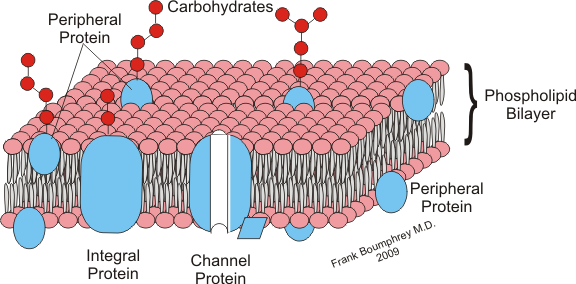
The cell membrane is formed out of they same stuff that vesicles are, however it is much larger and more complex. The primary function of the cell membrane is to contain the cell and to stop its organelles floating off. However it also regulates the flow of chemicals in an out of the cell and prevents crucial chemicals floating out. The cell membrane is made out of a bi-layer of fatty acids just as vesicles are. the fatty acids have a hydrophobic end and a hydrophilic end. The hydrophobic ends join together in the middle to form a bi-layer.
 |
| A diagram of the cell membrane. Note the double layer of fatty acids. Also note the channel proteins that are used in controlling the movement of materials in and out of the cell, through the membrane. http://upload.wikimedia.org/wikipedia/commons/f/fd/Cell_membrane3.png |
In the next part you can look forward to learning about the extra organelles in plant cells
Friday 27 April 2012
The Human Skeleton
(Key words are highlighted)
There are three main functions of the skeleton:
Support: The skeleton is what holds your body up right, without it we would be jelly!
Protection: The skeleton protects your inner organs for example your rib cage protects a lot of your internal organs such as the heart, liver, lungs, stomach and kidneys.
Movement: The skeleton allows you to do complex movements such as walking, running and lifting objects (along with your muscles).
The human skeleton is mainly made up of long bones as well as cartilage and various other different bones. below is a picture of the main structure of a typical long bone:
 Blood Vessels: The bone contains blood vessels as it needs blood to stop it from dying as it is alive, also the bone marrow creates new red blood cells thus it needs to pass the new blood cells into the blood stream.
Bone Marrow: The bone marrow is in the inside the middle of the bone, it is what creates the new red blood cells as they cannot self replicate as they lack a nucleus.
Hard Bone: Hard bone is what gives the bone structure and support as well as protecting the bone marrow.
Cartilage: Cartilage is very important, it is what stops your bones from rubbing and smashing together. Cartilage is found at the head of a bone where the joints are to absorb shock. If there wasn't any cartilage to absorb the shock then your bones, even from the smallest bit of shock forced upon the bones, could receive extreme harm and potential cracking or fracturing. Cartilage can get worn away quite quickly and the older your are the less quickly it regrows.
Air Pockets: Air pockets keep the bone light and flexible, without air pockets you would weigh significantly more and your bones wouldn't be able to take as much stress/strain.
There are three main functions of the skeleton:
Support: The skeleton is what holds your body up right, without it we would be jelly!
Protection: The skeleton protects your inner organs for example your rib cage protects a lot of your internal organs such as the heart, liver, lungs, stomach and kidneys.
Movement: The skeleton allows you to do complex movements such as walking, running and lifting objects (along with your muscles).
The human skeleton is mainly made up of long bones as well as cartilage and various other different bones. below is a picture of the main structure of a typical long bone:

Bone Marrow: The bone marrow is in the inside the middle of the bone, it is what creates the new red blood cells as they cannot self replicate as they lack a nucleus.
Hard Bone: Hard bone is what gives the bone structure and support as well as protecting the bone marrow.
Cartilage: Cartilage is very important, it is what stops your bones from rubbing and smashing together. Cartilage is found at the head of a bone where the joints are to absorb shock. If there wasn't any cartilage to absorb the shock then your bones, even from the smallest bit of shock forced upon the bones, could receive extreme harm and potential cracking or fracturing. Cartilage can get worn away quite quickly and the older your are the less quickly it regrows.
Air Pockets: Air pockets keep the bone light and flexible, without air pockets you would weigh significantly more and your bones wouldn't be able to take as much stress/strain.
Tuesday 24 April 2012
Topics on The Human Body
The Human Body
Here are the topics that we will be discussing over the next few weeks:The Skeleton - purpose and structure
Cellular Biology - internal workings of a cell
Muscles - functions
Here are just a few of them. Post us your ideas so we can go through what you want to know. We will be adding more information that isn't mentioned here so don't panic!! Please also post feedback in the comments as well as any additional information you would like to know. Thank you.Toby and Mike
Subscribe to:
Posts (Atom)